Medical & affiliate disclosure: CTX Stat provides educational laboratory information and is not a substitute for professional medical advice, diagnosis, or treatment. CTX Stat may receive compensation from some outbound provider links when an affiliate program is active; provider comparisons and rankings are based on consumer fit, effective cost, access, policies, and reputation—not commission.
A D-dimer blood test measures a small protein fragment that appears in the blood when the body forms and breaks down a fibrin blood clot. Clinicians most often use the test as part of an evaluation for venous thromboembolism—deep vein thrombosis (DVT) or pulmonary embolism (PE)—and sometimes as part of a workup for serious clotting activation such as disseminated intravascular coagulation (DIC).
The key point is that D-dimer is usually more useful when it is normal than when it is high. In the right clinical setting, a negative D-dimer can help rule out a dangerous clot and may prevent unnecessary imaging. A high D-dimer, however, does not diagnose a clot by itself because many conditions can raise it, including infection, inflammation, pregnancy, cancer, liver disease, heart disease, older age, trauma, recent surgery, and hospitalization. MedlinePlus summarizes this practical interpretation: low or normal levels make a clotting disorder less likely, while higher-than-normal levels require clinical context and often additional testing, not automatic diagnosis. MedlinePlus: D-dimer test
Quick take
- What it measures: D-dimer, a fibrin breakdown product released when clots are being dissolved.
- Common uses: Helping rule out DVT or PE in people with low or intermediate clinical probability; supporting evaluation of DIC in very ill patients.
- Preparation: Usually none. It is a standard blood draw from a vein.
- Most common cutoff: Many laboratories use a cutoff around 0.50 mg/L FEU, 0.5 mcg/mL FEU, or 500 ng/mL FEU, but units and assay methods vary.
- Important limitation: A high result is nonspecific. It tells your clinician that clot breakdown is increased, not where the clot is or why the level is elevated.
- Emergency note: New chest pain, shortness of breath, coughing blood, fainting, or sudden one-sided leg swelling should be treated as urgent symptoms, not as a reason to wait for an outpatient lab result.
What the D-dimer test measures
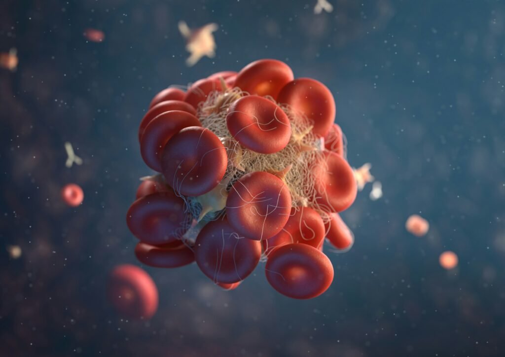
To understand D-dimer, it helps to know the basics of clot formation. When bleeding or blood-vessel injury occurs, the body uses a clotting protein called fibrin to stabilize the clot. Later, as the clot is remodeled or broken down, fragments of cross-linked fibrin enter the bloodstream. One of those fragments is D-dimer.
That means D-dimer is not a direct picture of a clot. It is a marker that the body has recently made and broken down fibrin. If a large clot is forming or dissolving, D-dimer can rise. But D-dimer can also rise whenever clotting and inflammation are activated throughout the body, which is why the test must be interpreted with symptoms, physical exam findings, risk factors, and sometimes imaging.
| Feature | What it means for consumers |
|---|---|
| Test type | Blood test, usually from a vein in the arm |
| Main analyte | D-dimer, a fibrin breakdown product |
| Most common clinical role | Helping rule out DVT or PE when clinical probability is not high |
| Diagnostic by itself? | No. A positive result does not prove a blood clot. |
| Special preparation | Usually none |
When a D-dimer blood test is ordered
A clinician may order a D-dimer blood test when symptoms and risk factors raise concern for abnormal clotting, especially DVT or PE. DVT is a clot in a deep vein, most often in the leg. PE occurs when clot material travels to the lungs and blocks blood flow. Mayo Clinic notes that D-dimer testing is one of several tests used in DVT evaluation, while duplex ultrasound is commonly used to diagnose DVT when imaging is needed. Mayo Clinic: DVT diagnosis and treatment
Typical reasons a clinician may consider D-dimer testing include:
- Possible DVT: one-sided leg swelling, calf or thigh pain, warmth, tenderness, or color change, especially after immobility, surgery, injury, pregnancy, estrogen therapy, cancer, or a prior clot.
- Possible PE: unexplained shortness of breath, chest pain that may worsen with breathing, rapid heart rate, fainting, low oxygen levels, or coughing blood.
- Possible DIC: a severe systemic clotting disorder usually evaluated in hospitals, often in the setting of sepsis, trauma, cancer, obstetric emergencies, or major illness.
- Monitoring in selected situations: D-dimer may be followed over time in some hospitalized or specialist-managed settings, but it is not a general wellness screening test.
Professional guidelines emphasize that D-dimer works best as part of a diagnostic strategy, not as a stand-alone screening test. The American Society of Hematology recommends D-dimer-based strategies for excluding PE in low-pretest-probability populations and recommends against using a positive D-dimer alone to diagnose PE. The same guideline notes that age-adjusted cutoffs may be used in appropriate adults when the assay cutoff is 500 µg/L. American Society of Hematology VTE diagnosis guideline
When D-dimer may not be the right first test
D-dimer is not always helpful. If a person has a high probability of DVT or PE based on symptoms, risk factors, and examination, clinicians often proceed directly to imaging rather than using D-dimer to decide. Likewise, hospitalized patients, people soon after surgery, people with active cancer, pregnant patients, and people with serious infections often have elevated D-dimer even without a new DVT or PE. In these groups, a positive result may add little clarity.
ARUP Consult, a laboratory medicine resource, notes that D-dimer should be used as an initial test only when results will be timely and when patients do not have other underlying conditions expected to cause baseline elevation. ARUP Consult: venous thromboembolism testing
Preparation: what to do before the test
Most people do not need to fast, stop medicines, or change activities before a D-dimer blood test. MedlinePlus states that no special preparation is needed for the D-dimer test. MedlinePlus: D-dimer test preparation
Even though preparation is simple, there are a few practical steps that can improve interpretation:
- Tell the ordering clinician about blood thinners. Anticoagulants can affect clot activity and may influence D-dimer interpretation. Do not stop a prescribed blood thinner unless your clinician specifically tells you to.
- Share recent events. Recent surgery, hospitalization, trauma, childbirth, infection, long travel, cancer treatment, or a previous clot can change the meaning of the result.
- Bring symptom timing. When symptoms started matters. A clinician interpreting possible DVT or PE will want to know whether symptoms began hours, days, or weeks ago.
- Use the same units when comparing results. D-dimer reports may use FEU or DDU units, and the numbers are not directly interchangeable without conversion.
What happens during the blood draw
The sample is usually collected from a vein in the arm. A health professional cleans the skin, places a small needle into a vein, collects blood into a tube, and removes the needle. The draw itself often takes only a few minutes. Mild bruising, soreness, or a small amount of bleeding can occur at the needle site, but serious complications from routine venipuncture are uncommon.
Turnaround time depends on where the test is performed. In emergency departments and hospitals, D-dimer can often be processed quickly because decisions about imaging or treatment may be time-sensitive. Outpatient reference-lab testing may take longer, especially if the sample must be transported.
How to interpret D-dimer results
D-dimer results are usually reported as negativeormal or positive/elevated, often with a numeric value and a cutoff. Interpretation depends on three things:
- Your clinical probability: how likely a clot seems based on symptoms, exam, and risk factors.
- The assay and units: FEU and DDU are different reporting systems.
- The cutoff used: standard, age-adjusted, or another validated diagnostic algorithm.
| Result pattern | Common meaning | What it does not mean |
|---|---|---|
| Normal or negative D-dimer | In a low- or intermediate-risk person, it can make DVT or PE unlikely enough that imaging may not be needed. | It does not rule out every clot in every situation, especially if clinical probability is high or testing conditions are not appropriate. |
| Mildly elevated D-dimer | Clot breakdown is increased or the cutoff has been exceeded. | It does not prove DVT, PE, stroke, heart attack, or DIC. |
| Markedly elevated D-dimer | May be seen with significant clot burden, severe infection, inflammation, trauma, cancer, DIC, or other serious illness. | The number alone does not locate a clot or identify the cause. |
| Falling D-dimer in monitored illness | May suggest decreasing clot formation/breakdown in a treated condition, depending on context. | It does not automatically mean the underlying condition is fully resolved. |
Common units: FEU vs DDU
One of the most confusing parts of D-dimer interpretation is the unit system. Many U.S. laboratories report D-dimer in fibrinogen equivalent units (FEU), while others use D-dimer units (DDU). Quest Diagnostics explains that 1.0 µg/mL FEU is approximately equivalent to 0.5 µg/mL DDU, so comparing numbers from different labs without checking units can lead to mistakes. Quest also states that its D-dimer cutoff is 0.5 µg/mL FEU and emphasizes that a thromboembolic event should not be excluded solely on the basis of the D-dimer level. Quest Diagnostics: D-dimer quantitative FAQ
| Reporting format | Approximate common cutoff example | Notes |
|---|---|---|
| ng/mL FEU | 500 ng/mL FEU | Same scale as 0.50 mg/L FEU or 0.5 mcg/mL FEU |
| mg/L FEU | 0.50 mg/L FEU | Commonly used by large laboratories |
| mcg/mL FEU | 0.5 mcg/mL FEU | Equivalent to 0.50 mg/L FEU |
| DDU | Often about half the FEU value | Do not compare directly with FEU without checking the lab’s reference information |
Age-adjusted D-dimer
D-dimer tends to rise with age, which reduces the test’s specificity in older adults. In some adult patients over age 50 with low or intermediate clinical probability of PE, clinicians may use an age-adjusted cutoff. The 2026 AHA/ACC/ACCP/ACEP/CHEST and collaborating-society guideline for acute pulmonary embolism states that, for fibrinogen equivalent unit assays, an age-adjusted D-dimer below age × 10 µg/L can effectively exclude PE in adults with low or intermediate clinical probability. 2026 guideline for acute pulmonary embolism in adults
For example, under that method, a 70-year-old might have an age-adjusted FEU threshold of 700 µg/L rather than the standard 500 µg/L. This does not mean older adults can self-adjust results at home. Age-adjusted interpretation is intended for specific clinical algorithms and compatible assays.
Why D-dimer can be high without a blood clot diagnosis
D-dimer is sensitive to clot formation and breakdown, but it is not specific for DVT or PE. A positive result may occur when the body is inflamed, healing, fighting infection, recovering from injury, or responding to cancer or surgery.
| Situation | Why D-dimer may rise |
|---|---|
| Recent surgery or trauma | Tissue injury and healing activate coagulation and fibrin turnover. |
| Pregnancy and the postpartum period | Coagulation activity changes naturally, and standard cutoffs may be less useful. |
| Infection or sepsis | Inflammation and coagulation pathways interact, sometimes strongly. |
| Cancer | Some cancers increase clotting tendency and baseline D-dimer. |
| Liver disease | Coagulation balance and clearance of clotting-related proteins may be altered. |
| Older age | Baseline levels are more likely to exceed standard cutoffs. |
| Rheumatoid arthritis and other inflammatory disorders | Systemic inflammation can increase fibrin turnover. |
D-dimer can also be misleadingly low in some circumstances. Testing too late after symptoms begin, a small clot burden, sample or assay issues, or being on anticoagulant therapy may affect results. This is one reason a clinician may order imaging despite a D-dimer result if the overall picture remains concerning.
D-dimer and DIC
DIC is a serious disorder in which clotting becomes abnormally activated throughout the body. It can cause both clotting and bleeding because platelets and clotting factors are consumed. D-dimer is one of several tests used in DIC evaluation, along with platelet count, prothrombin time (PT), partial thromboplastin time (PTT or aPTT), fibrinogen, and clinical assessment. The Merck Manual Professional Edition describes DIC diagnosis as using platelet count, PT, PTT, fibrinogen, plasma D-dimer, and a blood smear when DIC is suspected. Merck Manual Professional: disseminated intravascular coagulation
For consumers, the practical takeaway is that D-dimer alone does not diagnose DIC. DIC is usually evaluated in hospital settings because it is tied to severe underlying illness and requires urgent management of the cause.
What happens after an abnormal D-dimer result?
The next step depends on symptoms, risk level, and why the test was ordered. A positive D-dimer may lead to imaging, repeat evaluation, or additional blood tests. It should not be interpreted in isolation.
| Clinical question | Common next test or action | Purpose |
|---|---|---|
| Possible DVT in the leg | Compression or duplex ultrasound | Looks for impaired blood flow or visible clot in deep veins |
| Possible PE | CT pulmonary angiography or ventilation-perfusion scan, depending on the patient and setting | Evaluates blood flow and clot in the lungs |
| Possible DIC | Platelet count, PT/INR, aPTT, fibrinogen, blood smear, repeat labs | Assesses widespread clotting activation and factor consumption |
| High D-dimer without clot symptoms | Clinical review and targeted testing based on history | Looks for infection, inflammation, liver disease, cancer-related issues, recent injury, or other causes when appropriate |
If the D-dimer was ordered during an urgent evaluation, follow the instructions given by the treating clinician or emergency department. If the test was ordered outpatient and the result is elevated, contact the ordering clinician promptly for interpretation. Do not start aspirin, anticoagulants, supplements, or other treatments for a suspected clot unless a clinician has advised you to do so.
When to seek urgent care
A D-dimer test should not delay emergency evaluation for symptoms that could represent PE, DVT, stroke, heart attack, or severe bleeding. Seek urgent medical care or call emergency services for:
- Sudden or worsening shortness of breath
- Chest pain, especially if sharp, severe, or worse with breathing
- Coughing blood
- Fainting, severe dizziness, or blue lips
- One-sided leg swelling with pain, warmth, or redness
- New neurologic symptoms such as facial droop, arm weakness, trouble speaking, or sudden severe headache
- Uncontrolled bleeding, black stools, vomiting blood, or signs of shock
D-dimer blood test cost and access
The cost of a D-dimer blood test depends on where it is performed and why it is ordered. In an emergency department, the lab charge is usually only one part of a larger bill that may include facility fees, clinician evaluation, imaging, medications, and observation. In an outpatient setting, a clinician-ordered D-dimer may be billed through insurance or paid as a self-pay laboratory test.
When comparing self-pay options, look at the effective total cost, not just the advertised test price. The total may include:
- the D-dimer assay price;
- blood draw or specimen collection fees;
- clinician order or telehealth review fees if required;
- processing or administrative fees;
- state restrictions or limitations on direct-access testing;
- whether the advertised price includes a result report, clinician interpretation, or only the lab value.
For a potentially urgent clot concern, the least expensive outpatient lab option may not be the safest option if symptoms require immediate evaluation. D-dimer is most useful when integrated with clinical probability scoring and a plan for imaging if needed. If you are comparing prices for a nonemergency, clinician-approved outpatient test, confirm the collection site, turnaround time, all required fees, and whether your state allows the ordering workflow being offered.
Questions to ask about your result
If you receive a D-dimer result and are unsure what it means, these questions can help you have a more productive conversation with your clinician:
- Was my pretest probability considered low, intermediate, or high?
- What units did this lab use—FEU or DDU?
- Was an age-adjusted or standard cutoff applied?
- Could pregnancy, recent surgery, infection, inflammation, cancer, liver disease, or a recent injury explain this value?
- Do I need imaging, repeat testing, or no further testing?
- If I am on a blood thinner, how does that affect interpretation?
- Which symptoms should prompt emergency care?
FAQs about the D-dimer blood test
Is a D-dimer blood test a blood clot test?
It is a blood test related to clot formation and breakdown, but it is not a definitive “blood clot test.” A normal D-dimer can help rule out DVT or PE in selected low- or intermediate-risk patients. A high D-dimer suggests increased fibrin breakdown but does not prove that a clot is present.
What is a normal D-dimer result?
Many laboratories use a cutoff around 0.50 mg/L FEU, 0.5 mcg/mL FEU, or 500 ng/mL FEU, but reference information depends on the assay and units. Some situations use age-adjusted or algorithm-specific thresholds. Always read the result with the lab’s reference range and the ordering clinician’s interpretation.
Does a high D-dimer mean I have a pulmonary embolism?
No. A high D-dimer can occur with PE, but it can also occur with many non-PE conditions such as infection, inflammation, recent surgery, trauma, pregnancy, cancer, liver disease, and older age. PE diagnosis usually requires clinical assessment and, when indicated, imaging such as CT pulmonary angiography or a V/Q scan.
Can a normal D-dimer miss a clot?
It can, especially if the test is used outside the right clinical context. D-dimer is most reliable for ruling out DVT or PE when pretest probability is low or intermediate and a sensitive assay is used correctly. If clinical suspicion is high, clinicians may order imaging even if a D-dimer is normal.
Do I need to fast for a D-dimer test?
Usually no. Most people do not need to fast or make special preparations. Follow any instructions from the ordering clinician or laboratory, especially if other tests are being drawn at the same time.
How long does it take to get D-dimer results?
In hospitals and emergency departments, results may be available relatively quickly because care decisions may depend on them. Outpatient turnaround varies by collection site, laboratory, transport schedule, and ordering workflow.
Can pregnancy raise D-dimer?
Yes. D-dimer commonly rises during pregnancy, and standard cutoffs may be less useful. Pregnant patients with symptoms of DVT or PE need prompt clinical evaluation using pregnancy-appropriate diagnostic pathways.
Can COVID-19 or another infection raise D-dimer?
Yes. Significant infections and systemic inflammation can raise D-dimer because inflammation and coagulation pathways are connected. A high D-dimer during infection does not automatically mean a person has DVT or PE, but it may influence clinical evaluation depending on symptoms and severity.
Should I order a D-dimer test on my own?
D-dimer is usually not a good stand-alone screening test because positive results are common and nonspecific. If you have symptoms that could suggest a clot, seek medical evaluation rather than relying on a self-ordered lab. If a clinician recommends outpatient testing, make sure you understand what result would trigger follow-up.
What is the difference between D-dimer, PT/INR, aPTT, and fibrinogen?
D-dimer measures a fibrin breakdown product. PT/INR and aPTT measure how long blood takes to clot through different coagulation pathways. Fibrinogen measures a clotting protein that helps form fibrin. In conditions such as DIC, clinicians often interpret these tests together rather than relying on D-dimer alone.
Bottom line
The D-dimer blood test is a useful, fast marker of clot breakdown, especially for ruling out DVT or pulmonary embolism in the right patients. Its biggest limitation is that many common medical situations can elevate it. A high D-dimer should be treated as a clue that needs context, not a diagnosis. The safest interpretation comes from combining the lab result with symptoms, risk factors, timing, medications, units, and—when needed—imaging or additional blood tests.
Source Notes
- MedlinePlus: D-dimer test preparation
- Mayo Clinic: Deep Vein Thrombosis Diagnosis and Treatment
- American Society of Hematology 2018 Guidelines for Management of Venous Thromboembolism: Diagnosis of VTE
- ARUP Consult: venous thromboembolism testing
- Quest Diagnostics: D-dimer quantitative FAQ
- 2026 Guideline for the Evaluation and Management of Acute Pulmonary Embolism in Adults
- Merck Manual Professional Edition: Disseminated Intravascular Coagulation